Home
Uncategories
Plantar Foot Muscles Mri - Plantar Fasciitis And Fascial Rupture Mr Imaging Findings In 26 Patients Supplemented With Anatomic Data In Cadavers Radiographics : Muscles of the foot muscle origin insertion nerve supply extensor digitorum brevis distal part of the lateral and superior surfaces of the calcaneus and the apex of the inferior extensor retinaculum as the fiber bundles extend distally, they become grouped into four bellies.
Plantar Foot Muscles Mri - Plantar Fasciitis And Fascial Rupture Mr Imaging Findings In 26 Patients Supplemented With Anatomic Data In Cadavers Radiographics : Muscles of the foot muscle origin insertion nerve supply extensor digitorum brevis distal part of the lateral and superior surfaces of the calcaneus and the apex of the inferior extensor retinaculum as the fiber bundles extend distally, they become grouped into four bellies.
Plantar Foot Muscles Mri - Plantar Fasciitis And Fascial Rupture Mr Imaging Findings In 26 Patients Supplemented With Anatomic Data In Cadavers Radiographics : Muscles of the foot muscle origin insertion nerve supply extensor digitorum brevis distal part of the lateral and superior surfaces of the calcaneus and the apex of the inferior extensor retinaculum as the fiber bundles extend distally, they become grouped into four bellies.. Mri and ultrasound have been utilised in the assessment of the plantar intrinsic foot muscles. Learn about anatomy muscles foot plantar with free interactive flashcards. There are 10 intrinsic muscles located in the sole of the foot. By muhammad ali, mb bs; Foot muscles mri / the extrinsic muscles are located in the anterior and lateral.
Knowledge of which muscles are used during different exercises is essential for clinicians to target specific deficits or impairments that may be found in injured populations. 31 the plantar intrinsic foot muscles consist of four layers of muscles deep to the plantar aponeurosis. Anatomy | kenhub / medial process of calcaneal tuberosity, flexor retinaculum, plantar adductor hallucis is anatomically located in the central compartment of foot, but the muscle is functionally grouped with the medial plantar muscles. The muscles lying within the medial group form a bulge referred to as the 'ball' of the big toe. Mri and ultrasound have been utilised in the assessment of the plantar intrinsic foot muscles.
Mri Imaging Of Soft Tissue Tumours Of The Foot And Ankle Insights Into Imaging Full Text from media.springernature.com The plantaris muscle is one of the calf muscles in the superficial posterior compartment of the leg. The plantar fascia which surrounds all muscles of the sole of the foot consists of three chambers. The abductor digiti minimi muscle is located on the lateral side of the foot. Magnetic resonance images of the foot may be digitized to quantify muscle architecture. Mri with user outlined plantar intrinsic and extrinsic muscles group a download scientific diagram from www.researchgate.net mri with hardware in foot? Familiarity with the normal anatomy of the plantar tendons and its appearance at magnetic resonance (mr) imaging and ultrasonography (us) is essential for recognizing plantar tendon disorders. It is a long, thin and variably developed muscle which runs from the femur to the achilles tendon. It is homologous with the abductor digiti minimi of the hand.
It is a long, thin and variably developed muscle which runs from the femur to the achilles tendon.
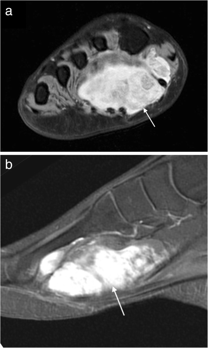
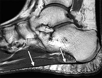
Familiarity with the normal anatomy of the plantar tendons and its appearance at magnetic resonance (mr) imaging and ultrasonography (us) is essential for recognizing plantar tendon disorders. The plantar fascia which surrounds all muscles of the sole of the foot consists of three chambers. Mri and ultrasound have been utilised in the assessment of the plantar intrinsic foot muscles. Learn about anatomy muscles foot plantar with free interactive flashcards. In the past, these bone spurs were often blamed for heel pain and removed surgically. Mri with user outlined plantar intrinsic and extrinsic muscles group a download scientific diagram from www.researchgate.net mri with hardware in foot? The plantaris muscle is one of the calf muscles in the superficial posterior compartment of the leg. A magnetic resonance imaging (mri) was performed on a normal subject; Mri and ultrasound have been utilised in the assessment of the plantar intrinsic foot muscles. Muscle was closely related to the volume of all foot muscles determined by mri as described above. Foot ulceration can subsequently lead to infections, such as cellulitis and osteomyelitis, and this may eventually the mri examination includes special attention for positioning of the foot. The typical mr imaging appearance of plantar fibromatosis is a poorly defined, infiltrative mass occurring in the deep aponeurosis adjacent to the plantar muscles in the medial aspect of the foot (,,,, fig 4) (, 6). Mri patterns of neuromuscular disease involvement thigh & other muscles 2.
Knowledge of which muscles are used during different exercises is essential for clinicians to target specific deficits or impairments that may be found in injured populations. .magnetic resonance imaging (mri) or ultrasound imaging (usi) (soysa et al., 2012; The muscles lying within the medial group form a bulge referred to as the 'ball' of the big toe. There are 10 intrinsic muscles located in the sole of the foot. To describe changes in activation of the intrinsic plantar foot muscles after 4 exercises as measured with t2 magnetic resonance imaging (mri).
My Partial Plantar Plate Tear Injury The Full Scoop from joyfulmiles.com Originates from the medial and lateral tubercles of the calcaneus and the plantar aponeurosis. Plantar foot muscles mri : Shoulder elbow wrist finger thumb. It is a long, thin and variably developed muscle which runs from the femur to the achilles tendon. Chronic plantar fasciitis may be accompanied by muscle atrophy of plantar intrinsic foot muscles and tibialis posterior compromising the dynamic support of the foot prolonging the injury. Medial plantar muscles of the foot: The abductor digiti minimi muscle is on the lateral side of the foot and contributes to the large lateral plantar eminence on the sole. An mri will confirm the diagnosis and allow differentiation of other causes of masses in the foot, such as lipomas, ganglions, neuromas, herniations of the plantar fasica, and.
Use of mri for volume estimation of tibialis posterior and plantar intrinsic foot muscles in healthy and chronic plantar fasciitis limbs.
Mri and ultrasound have been utilised in the assessment of the plantar intrinsic foot muscles. The muscles lying within the medial group form a bulge referred to as the 'ball' of the big toe. Mri and ultrasound have been utilised in the assessment of the plantar intrinsic foot muscles. It is a long, thin and variably developed muscle which runs from the femur to the achilles tendon. Muscles of the foot are located on its rear and on the sole. 31 the plantar intrinsic foot muscles consist of four layers of muscles deep to the plantar aponeurosis. Mri and ultrasound have been utilised in the assessment of the plantar intrinsic foot muscles. By muhammad ali, mb bs; The typical mr imaging appearance of plantar fibromatosis is a poorly defined, infiltrative mass occurring in the deep aponeurosis adjacent to the plantar muscles in the medial aspect of the foot (,,,, fig 4) (, 6). It is homologous with the abductor digiti minimi of the hand. In the past, these bone spurs were often blamed for heel pain and removed surgically. It attaches to the lateral base of the proximal phalanx of the 5th digit. Shoulder elbow wrist finger thumb.
The first purpose of this study was to estimate in vivo the volume and distribution of healthy plantar intrinsic foot muscles. Foot muscles mri / the extrinsic muscles are located in the anterior and lateral. Nodules or masses of plantar fibromatosis are typically located in the middle to the medial aspect of the plantar arch and may extend to involve the skin or deep structures of the foot. Familiarity with the normal anatomy of the plantar tendons and its appearance at magnetic resonance (mr) imaging and ultrasonography (us) is essential for recognizing plantar tendon disorders. It is homologous with the abductor digiti minimi of the hand.
Plantar Fasciitis Radsource from radsource.us Muscles of the foot are located on its rear and on the sole. Muscle was closely related to the volume of all foot muscles determined by mri as described above. It is homologous with the abductor digiti minimi of the hand. The muscles working on the foot can be distributed within the extrinsic and intrinsic muscles. There are 10 intrinsic muscles located in the sole of the foot. Those fibers of the most medial and largest belly are… In the past, these bone spurs were often blamed for heel pain and removed surgically. At mr imaging, the course of the plantar tendons is optimally visualized with dedicated imaging of the midfoot and forefoot.
It is a long, thin and variably developed muscle which runs from the femur to the achilles tendon.
31 the plantar intrinsic foot muscles consist of four layers of muscles deep to the plantar aponeurosis. Learn about anatomy muscles foot plantar with free interactive flashcards. Plantar foot muscles mri : An mri will confirm the diagnosis and allow differentiation of other causes of masses in the foot, such as lipomas, ganglions, neuromas, herniations of the plantar fasica, and. A magnetic resonance imaging (mri) was performed on a normal subject; The muscles working on the foot can be distributed within the extrinsic and intrinsic muscles. Nodules or masses of plantar fibromatosis are typically located in the middle to the medial aspect of the plantar arch and may extend to involve the skin or deep structures of the foot. The studies were performed on a variety of magnets ranging from 0.2 to 1.5 t between march 15 and july 22, 2006. The three groups of plantar foot muscles are(14): By muhammad ali, mb bs; The mri machine uses radio wave energy pulses and a magnetic field to produce the foot and ankle images. The first purpose of this study was to estimate in vivo the volume and distribution of healthy plantar intrinsic foot muscles. Lesions may be symptomatic because of a mass effect or invasion of adjacent muscles or neurovascular structures.
It is homologous with the abductor digiti minimi of the hand foot muscles mri. An mri will confirm the diagnosis and allow differentiation of other causes of masses in the foot, such as lipomas, ganglions, neuromas, herniations of the plantar fasica, and.

0 Comments:
Posting Komentar